Nigerian AI Researcher Develops Technology That Could Reduce MRI Scan Times by 90%
Mary-Brenda Akoda’s award-winning innovation uses artificial intelligence to reconstruct medical images from less scan data, potentially helping hospitals improve diagnostic capacity and reduce waiting times.

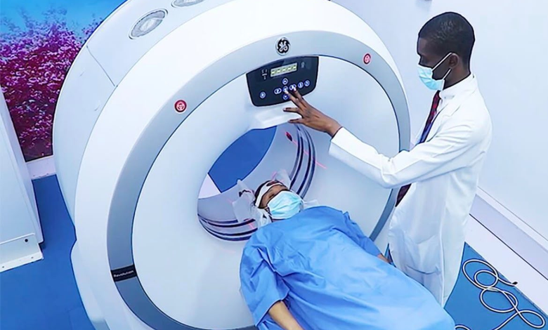
A Nigerian artificial intelligence researcher, Mary-Brenda Akoda, has developed a medical imaging technology designed to reduce Magnetic Resonance Imaging (MRI) scan times by up to 90 per cent, offering a potential solution to one of the challenges facing modern healthcare: the time and resources required to produce detailed diagnostic images.
The technology, known as GenScan AI or GenMRI, applies artificial intelligence to reconstruct MRI images using significantly less acquired data. By reducing the amount of information that must be collected during a scan, the system aims to produce diagnostic-quality images in a fraction of the usual time.
The innovation earned Akoda the 2026 Nigeria Prize for Science and Innovation, a $100,000 award recognising scientific solutions to major societal challenges. Her work places a spotlight on the growing contribution of African researchers to the development of technologies that address practical problems in healthcare.
How the technology works
MRI scanners use strong magnetic fields and radio waves to produce detailed images of structures inside the body. Doctors rely on these images to investigate conditions affecting the brain, spinal cord, joints, muscles and internal organs, among other parts of the body.
Despite their importance, MRI examinations can take considerable time, depending on the body part being examined, the imaging protocol and the level of detail required. Patients may need to remain still for extended periods, while hospitals must manage scanner availability, staff schedules and growing demand for diagnostic services.
GenMRI seeks to address this challenge through AI-powered image reconstruction. Rather than relying on the same volume of data traditionally collected during a scan, the system is designed to use a smaller amount of acquired information and computational methods to reconstruct the resulting images.
In practical terms, the technology aims to reduce the time patients spend inside the scanner while retaining the image quality required for medical interpretation.
Under the reported performance target, an MRI examination that takes approximately 20 minutes could potentially be reduced to around two minutes, representing a 90 per cent reduction in scanning time.
However, the actual time saved would depend on the type of examination, the scanner being used and the clinical requirements of each patient. The shorter scan time should therefore be understood as a reported potential rather than a guaranteed result for every procedure.
The ability to shorten MRI examinations could have implications for hospitals, diagnostic centres and patients, particularly in healthcare systems where access to advanced medical imaging remains limited.
Increased diagnostic capacity: If the technology can safely shorten examination times, hospitals could potentially scan more patients during existing operating hours. This could help facilities manage demand without immediately investing in additional scanners.
Reduced waiting times: Patients sometimes wait for appointments because diagnostic centres have limited scanning capacity. Faster examinations could help facilities process their appointment schedules more efficiently, although actual waiting times would also depend on staffing, demand and other operational constraints.
Improved patient experience: Remaining still inside an MRI scanner can be uncomfortable, especially for patients experiencing pain, anxiety or difficulty maintaining a fixed position. Shorter examinations could make the process more manageable for some patients.
More efficient use of expensive equipment: MRI machines require substantial investment, maintenance and specialist personnel. Improving the number of examinations a facility can perform with its existing equipment could help healthcare providers make better use of their resources.
These potential benefits are particularly relevant to countries where access to specialised diagnostic equipment is unevenly distributed between major cities and less-served communities.
Nevertheless, faster image acquisition alone cannot resolve every barrier to medical diagnosis. Equipment availability, electricity supply, maintenance, trained radiographers, specialist interpretation and the cost of examinations would remain important factors.
Akoda’s work has also been linked to a personal experience involving an elderly relative who suffered a stroke and needed an urgent MRI examination but encountered delays in accessing the scan.
The experience helped shape her interest in finding ways to make medical imaging faster and more accessible. It also illustrates how personal encounters with healthcare challenges can influence the direction of scientific research.
Rather than treating the problem solely as a question of purchasing more equipment, the technology explores whether artificial intelligence can help existing diagnostic infrastructure perform more efficiently.
This approach reflects a wider direction in medical technology, where researchers are developing AI systems to assist with image reconstruction, improve clinical workflows and support the delivery of diagnostic services.
From artificial intelligence research to medical applications
Akoda studied computer science at Goldsmiths, University of London, before completing a Master of Research in Artificial Intelligence and Machine Learning at Imperial College London. She has also worked as a software engineer and AI researcher at Microsoft.
Her background combines software engineering, machine learning and the development of computational systems fields increasingly being applied to healthcare.
AI-assisted MRI reconstruction is an established area of medical research internationally. Its central challenge is to produce useful images from less data without introducing distortions or concealing clinically important details.
That distinction is critical. A scan that takes less time is only valuable if the resulting images remain sufficiently reliable for the intended medical examination. A system must therefore be assessed not just on speed but also on image quality, consistency and performance across different patients, scanners and clinical settings.
Clinical validation and deployment remain important
GenScan AI has reported engagement with clinical partners in Nigeria, Kenya, the United Kingdom and the United States as it advances validation and prepares for pilot deployments. The company has identified the second half of 2027 as its target for wider deployment.
Before the technology can be adopted broadly, its performance will need to be established for the specific examinations and clinical settings in which it is intended to operate. This includes assessing whether reconstructed images preserve the details doctors need to identify abnormalities and make informed decisions.
Regulatory requirements, compatibility with hospital imaging systems, integration into clinical workflows and acceptance by radiologists will also influence how quickly the technology can be introduced into routine care.
The extent to which it can reduce costs or improve access will ultimately depend on these results, as well as the commercial model through which hospitals obtain and use the system.
Recognition for African scientific innovation
Akoda’s selection for the 2026 Nigeria Prize for Science and Innovation highlights the importance of research that connects advanced technology with practical societal needs.
Artificial intelligence is increasingly being explored across African industries, including agriculture, financial services, education and healthcare. Medical imaging presents a particularly important opportunity because delays in diagnosis can affect treatment decisions and the experience of patients navigating the healthcare system.
For African researchers, developing solutions suited to local healthcare challenges could also create opportunities for collaboration between universities, technology companies, hospitals and international research institutions.
If GenMRI demonstrates that it can consistently produce clinically reliable images while substantially reducing scan times, it could contribute to efforts to improve the efficiency of diagnostic services in Nigeria and other markets.
The next stage will be determining how well the technology performs under real clinical conditions and whether hospitals can implement it safely and sustainably.
For now, Akoda’s innovation represents a promising example of Nigerian-led research applying artificial intelligence to a significant healthcare challenge, with the potential to make advanced diagnostic services more efficient if clinical validation and deployment prove successful.
TNAM